
Mechanism underlying the immune checkpoint inhibitor-induced hyper-progressive state of cancer

Abstract
Immune checkpoint inhibitors (ICIs) are gradually replacing chemotherapy as the cornerstone of the treatment of advanced malignant tumors because of their long-lasting and significant effect in different tumor types and greatly prolonging the survival time of patients. However, not all patients can respond to ICIs, and even rapid tumor growth after treatment with ICI has been observed in a number of clinical studies. This rapid progression phenomenon is called hyper-progressive disease (HPD). The occurrence of HPD is not uncommon. Past statistics show that the incidence of HPD is 4%-29% in different tumor types, and the progression-free survival and overall survival of patients with HPD are significantly shorter than those of the non-HPD progressor group. With the deepening of the study of HPD, we have established a preliminary understanding of HPD, but the diagnostic criteria of HPD are still not unified, and the addition of biomarkers may break this dilemma. In addition, quite a few immune cells have been found to be involved in the occurrence and development of HPD in the tumor microenvironment, indicating that the molecular mechanism of HPD may be triggered by a variety of ongoing events at the same time. In this review, we summarize past findings, including case reports, clinical trials, and fundamental research; compare the diagnostic criteria, incidence, and clinical prognostic indicators of HPD in different studies; and explore the molecular mechanism and future research direction of HPD.
Keywords
INTRODUCTION
Immune checkpoint inhibitors (ICIs) have continuously promoted the progress of the treatment of malignant tumors since their advent. They have gradually replaced chemotherapy as the cornerstone for the treatment of malignant tumors; however, ICIs are only effective in some patients and remain ineffective in most populations. Changes in the tumor microenvironment (TME) induced by ICIs stimulate the accelerated growth of malignant tumor cells. This special tumor progression mode is called the hyper-progressive disease (HPD) state. Lahmar et al.[1] reported the HPD phenomenon for the first time in a wall newspaper at the 2016 European Society of Medical Oncology Annual Meeting. Eight patients with advanced non-small cell carcinoma (NSCLC) exhibiting fast progression at the time of initial examination were identified as HPD cases. HPD gained attention in 2017 when Champiat et al.[2] reported a 9% HPD incidence in 131 cancer patients in a phase I prospective study. Evidence of HPD, the phenomenon of early crossover of the survival curve, is also reported in some phase III clinical studies, including in NSCLC (CheckMate026[3], CheckMate057[4], and CheckMate227[5]), HNSCC (CheckMate141[6]), and uroepithelial carcinoma (Keynote045[7] and IMvigor211[8]). Patients receiving immunotherapy died at a greater rate in the first three months than those treated with chemotherapy. HPD is not unique to immunotherapy and can also be caused by chemotherapy[9] and targeted therapy[10]. However, the incidence of HPD after ICI treatment is significantly higher than in the chemotherapeutic regime[11]. Since its discovery in 2016, several studies on HPD have been reported in the last five years. Nevertheless, the incidence, diagnostic criteria, and pathogenesis of HPD remain in the preliminary stages. This review summarizes the recently published cases, clinical studies, and basic studies on HPD.
DIAGNOSTIC CRITERIA FOR HPD
At present, there is no agreement on the diagnostic criteria of HPD. Although many clinical studies on HPD adopt different diagnostic criteria, the diagnostic indicators of HPD mainly focus on the following five: tumor growth rate (TGR), ΔTGR, tumor growth kinetics (TGK), Response Evaluation Criteria in Solid Tumors (RECIST), and time to failure (TTF). TGR represents the percentage of monthly tumor volume growth (excluding new and immeasurable lesions), and the difference between the two at and before treatment is defined as ΔTGR. TGK is defined similarly to TGR, but it primarily reflects tumor growth rate per unit time. TTF refers to the time of treatment failure. Champiat et al.[2] earlier adopted such criteria as TGR > 2 and RECIST to assess the progress for the first time to define HPD. In the same year,
Although the volumetric method is superior to the RECIST standard, there are practical problems: first, not all patients can complete the pre-baseline computed tomography (CT) scan, especially those receiving ICI as late first-line treatment. Second, new and unmeasurable lesions cannot be measured by TGR.
Thus, combining indicators with each other may be more conducive to diagnosis. The radiological and clinical diagnostic criteria for HPD are still being explored. With the deepening of the understanding of biomarkers for HPD, biomarkers may be involved in the diagnostic criteria of HPD in the future, and the joint definition of HPD by three diagnostic methods may be more accurate and practical.
INCIDENCE AND PROGNOSTIC INDICATORS OF HPD
The incidence and clinical prognostic indicators of HPD are also different. Chen et al.[20] reviewed the medical records of 377 patients with multiple malignancies and reported the incidence of HPD (10.08%). Factors associated with HPD include the presence of more than two metastatic sites, Eastern Cooperative Oncology Group score ≥ 2, liver metastasis, and lactic dehydrogenase level higher than the normal upper limit. Kirsten rat sarcoma viral oncogene homolog status is significantly correlated with HPD in colon cancer patients. Two large-scale meta-analyses reported the incidence of HPD in patients with pan-cancer as 1%-30%[21] and 5.9%-43.1%[22]. The clinical prognostic markers used in these analyses were similar to those reported by Chen et al.[20]. Ferrara et al.[9], using RECIST 1.1 and TGR criteria, reported a 13.8% (56/406) HPD incidence in patients with advanced NSCLC; HPD was associated with more than two metastases before immunotherapy. Kim et al.[23] first defined three criteria (TGR, TGK, and TTF) to calculate the incidence of HPD (20.9%, 20.5%, and 37.3%, respectively). In HPD patients who satisfied both TGR and TGK criteria, poorer progression-free survival (PFS) and OS were observed. Although no clinicopathological variables of HPD were reported in the study, in the exploratory biomarker analysis of peripheral blood, CD8+ T lymphocytes, lower effector/memory subsets (CCR7-CD45RA- T cells in total CD8+ T cells), and higher populations of severely depleted cells (TIGIT+ T cells in PD-1+CD8+ T cells) were associated with HPD and poor survival. In two real-world studies, the incidence of HPD in advanced NSCLC was 19.2% (16/83)[24] and 8.1% (6/74)[25]. Among them, one study reported an increased rate of fluid accumulation (up to 90%) and decreased albumin level, while the other showed a significant increase in the number of circulating Treg cells in HPD patients. Chen et al.[26] performed a meta-analysis consisting of 1389 NSCLC patients from six clinical studies and found that the incidence of HPD was 8.02%-30.43%. The incidence of HPD and clinical prognostic indicators in cancer types are shown in Table 1.
Recent retrospective studies on hyper-progression after immunotherapy
| Tumor type | Agents | HPD criteria | HPD incidence | Prognostic indicators | Outcomes (HPD vs. non-HPD) | Ref. |
| Multiple tumor types | PD-1/PD-L1 inhibitor monotherapy | - | 1%-30% (217/1519) | Serum LDH > upper normal limit; > 2 metastatic sites prior to immunotherapy; liver metastatic sites; RMH prognostic score ≥ 2; positive PD-L1 expression status | - | Kim et al.[21] (2019) |
| Multiple tumor types | PD-1/PD-L1 inhibitor monotherapy | RECIST criteria (1.4× baseline sum target lesions or | RECIST criteria, 10.7% (29/270); TGR criteria, 6.3% (14/221) | RECIST criteria of no or TGR criteria of liver metastatic sites; > 2 metastatic sites prior to immunotherapy | OS: 5.23 months vs. 7.33 months, P = 0.04, by RECIST; 4.2 months vs. 6.27 months, P = 0.346, by TGR | Matos et al.[17] (2020) |
| Multiple tumor types | PD-1 inhibitors (nivolumab or pembrolizumab) | ΔTGR > 50% | 10.08% (38/377) | > 2 metastatic sites prior to immunotherapy; ECOG ≥ 2; hepatic metastases; serum LDH > upper normal limit; KRAS status in colorectal cancer | OS: 3.6 months vs. 7.3 months, P < 0.01 | Chen et al.[20] (2021) |
| Multiple tumor types | PD-1 or PD-L1 inhibitor monotherapy or combined with CTLA-4 inhibitor | 4 categories (TGR, TGK, early tumor burden increase, or combinations of the above) | 5.9%-43.1% (3109) | - | - | Park et al.[22] (2021) |
| NSCLC | PD-1 or PD-L1 inhibitor monotherapy or combined with CTLA-4 inhibitor | RECIST 1.1 progression and ΔTGR > 50% | 14% (56/406 treated with ICI); 5% (3/59 treated with chemotherapy) | > 2 metastatic sites prior to immunotherapy | OS: HR = 2.18, 95%CI: 1.29-3.69, P = 0.03 | Ferrara et al.[9] (2018) |
| NSCLC | PD-1 inhibitors (nivolumab) | < 3 nivolumab injections | 20% (57/292) | PS > 2 at nivolumab initiation | OS: 1.4 months vs. 13.5 months, P < 0.0001 | Costantini et al.[112] (2019) |
| NSCLC | PD-1 or PD-L1 inhibitor monotherapy | Volumetric time-dependent criteria (TGK ≥ 2) or one-dimensional criteria: RECIST 1.1 progression | 14.3% (48/335 by volumetric assessment); 13.1% (44/335 by one-dimensional criteria) | High neutrophil-to-lymphocyte ratio; LKB1 mutation | OS: 4.7 months vs. 7.9 months, P = 0.009, by volumetric; 5.2 months vs. 7.1 months, P = 0.288, by RECIST | Kim et al.[14] (2020) |
| NSCLC | PD-1 or PD-L1 inhibitor monotherapy | TGK ≥ 2, TGR ≥ 2, or TTF < 2 months | 20.9% (55/263 TGK), 20.5% (54/263 TGR), 37.3% (98/263 TTF) | ≥ 2 metastatic locations; liver metastases; neutrophils; neutrophil-to-lymphocyte ratio; LDH; high CD8+PD-1+TIGIT+ T cells; low CD8+CCR7-CD45RA- T cells | PFS: HR = 4.62, 95%CI: 2.87-7.44, P < 0.05; OS: HR = 5.71, 95%CI: 3.14-8.23, P < 0.05 | Kim et al.[23] (2019) |
| NSCLC | PD-1 inhibitors (nivolumab) | RECIST 1.1 progression and TGR ≥ 2 | 19.2% (16/83) | Pleura or pericardium metastasis; low circulating albumin | PFS: 0.43 months vs. 1.35 months; OS: 2.2 months vs. 4.1 months | Kim et al.[24] (2020) |
| NSCLC | PD-1 /PD-L1 inhibitor monotherapy or combined with other immunotherapy treatments | Ferté criteria (RECIST 1.1 progression and TGR ≥ 2), Le Tourneau criteria (TGK > 2), Garralda criteria (increase of ≥ 20% in target tumor burden plus multiple new lesions or increase of ≥ 40% in target tumor burden compared with baseline) or Caramella criteria (RECIST 1.1 progression and ΔTGR > 100%) | 5.4%-18.5% (406) | No (including previously described prognostic factors such as age, LDH, albumin, > 2 metastatic sites, RMH score) | - | Kas et al.[15] (2020) |
| NSCLC | PD-1 /PD-L1 inhibitor monotherapy or combined with other immunotherapy treatments | - | 8.02%-30.43% (1389) | ECOG > 1; RMH ≥ 2; serum LDH > upper Normal limit; > 2 metastatic sites prior to immunotherapy; liver metastases | - | Chen et al.[26] (2020) |
| NSCLC | PD-1 or PD-L1 inhibitor monotherapy or combined with CTLA-4 inhibitor | 5 definitions (TGR, ΔTGR, TGK, RECIST, or TTF) | 11.3%, 5.7%, 17%, 9.6%, 31.7% (169) | - | - | Abbar et al.[19] (2021) |
| NSCLC | PD-1 or PD-L1 inhibitor monotherapy | TGK > 2 and TTF ≤ 2 months | 11.3% (26/231) | Heavy smoker; PD-L1 expression ≤ 1%; ≥ 3 metastatic sites | OS: 5.5 months vs. 6.1 months | Kim et al.[110] (2021) |
| NSCLC | PD-1/PD-L1 inhibitor monotherapy or combined with chemotherapy | TGR > 2 | 17.6% (25/142 monotherapy); 2.9% (1/34 combination therapy) | - | - | Matsuo et al.[113] (2021) |
| NSCLC | PD-1 or PD-L1 inhibitor monotherapy | TGK ≥ 2 | 8.1% (6/74) | CD4+CD25+CD127loFoxP3+ Treg cells was increased on Day 7 after initiation of treatment | - | Kang et al.[25] (2021) |
| HNSCC | PD-1 or PD-L1 inhibitor monotherapy or combined with CTLA-4 inhibitor | TGK > 2 | 14.4% (18/125) | Younger age; primary tumor of oral cavity; previous locoregional irradiation | PFS: 1.2 months vs. 3.4 months, P < 0.001; OS: 3.4 months vs. 10.7 months, P = 0.047 | Park et al.[31] (2020) |
| HNSCC | PD-1 or PD-L1 inhibitor monotherapy or combined with CTLA-4 inhibitor | TGK ≥ 2 | 15.4% (18/117) | Primary site in the oral cavity; administration of ICI in the second/third setting | PFS: 1.8 months vs. 6.1 months, P = 0.0001; OS: 6.53 months vs. 15 months, P = 0.0018 | Economopoulou et al.[51] (2021) |
| MM | PD-1 inhibitor, CTLA-4 inhibitor monotherapy or combination | TTF < 2 months, doubling of tumor burden, and TGR > 2 | 1.3% (1/75) | - | - | Schuiveling et al.[114] (2021) |
| GC | PD-1 inhibitors (nivolumab) | TGK ≥ 2 and (SPOST/S0-1) > 0.5 | 22.1% (143) | PD-L1 CPS; MMR | PFS: 1.2 months vs. 1.7 months, P < 0.001; OS: 3.3 months vs. 6.8 months, P = 0.012 | Hagi et al.[115] (2020) |
| HCC | PD-1 inhibitors (nivolumab) | TGK > 4 and ΔTGR > 40% | 12.7% (24/189) | Neutrophil-to-lymphocyte ratio | PFS: HR = 2.194, 95%CI: 1.214-3.964; OS: HR = 2.238, 95%CI: 1.233-4.062 | Kim et al.[116] (2021) |
| RCC and UC | PD-1/PD-L1 inhibitor monotherapy | Tumor burden increase ≥ 50%, TGR ≥ 2, or ≥ 10 metastatic sites | 0.9% (1/102), 11.9% (12/101) | UC; creatinine > 1.2 mg/dL | PFS: 1.3 months vs. 3.9 months, P < 0.001; OS: 3.5 months vs. 7.3 months, P < 0.001 | Hwang et al.[117] (2020) |
| GYN | PD-1 inhibitor | Tumor burden increase of ≥ 40% or tumor burden increase of ≥ 20% plus multiple new lesions | 23.3% (14/60) | Neutrophil-to-lymphocyte ratio; > 3 metastatic sites | - | Rodriguez Freixinos et al.[118] (2018) |
CASE SUMMARY
The limitations of ICIs, as they may not be appropriate for some patients, caused “disease flare” in a 54-year-old man with stage IIB lung adenocarcinoma after 10th-line treatment with nivolumab[27]. This case opened up the HPD patient reports, and, according to incomplete statistical data, in 44 cases involving 53 patients, malignant tumor types were mainly distributed in the respiratory system, digestive system, and urinary system and were immune to single and double drugs to a significantly higher degree than due to the immune or anti-angiogenesis drugs with combination chemotherapy. Most patients with HPD after ICI treatment developed liver, lung, and brain metastases. Selected case studies are listed in Table 2. Among them, the youngest patient was a 13-year-old girl suffering from malignant melanoma, which progressed to HPD mode after two cycles of treatment with avelumab in palliative radiotherapy. The Food and Drug Administration has approved ICIs for the treatment of children with microsatellite unstable malignant tumors based on reports in adults[28]. However, the interaction between children’s immune systems and anti-PD1 therapy remains unclear. The oldest patient was an 80-year-old patient with lung squamous carcinoma[29]. The symptoms of HPD were pneumonia, pleural effusion, and pericardial effusion. Many patients developed the same symptoms after ICI treatment for malignant tumors of the respiratory system and digestive system and malignant melanoma. A previous study in South Korea reported a higher frequency of increased fluid accumulation in HPD patients with pleural or pericardial metastases after treatment with nivolumab as compared to the progressive disease (PD) patients without HPD [90% (9/10) vs. 28.6% (4/14); P = 0.005]; the circulating albumin level was significantly reduced in HPD patients (P = 0.030)[24]. A considerable proportion of HPD occurred in patients after radiotherapy, which suggested that radiotherapy had a bidirectional regulatory effect on the anti-tumor immune response. If the immunosuppressive function of radiotherapy is dominant, a combination of ICIs may lead to HPD[30]. A clinical study of head and neck squamous cell carcinoma also suggested that previous local irradiation was an important predictor of HPD[31]. In addition to being associated with radiotherapy, AKT1 E17K mutation[32] and PI3K/AKT pathway[33] were also related to HPD. Interestingly, after immunohistochemical staining of the primary tumor and metastases samples with HPD, Barham et al.[34] showed that the tumor infiltrating lymphocyte (TIL) number was not necessarily correlated with ICI response, as levels of granzyme B and TIA-1 of infiltrated CD8+ T cells were mostly negative, indicating that these were inflammatory T cells which cause tumor drug resistance and myocarditis. They cannot effectively dissolve the tumor, so additional functional markers are required to distinguish between inflammatory and cytolytic CD8+ TIL. For treatment, the salvage therapy in HPD has not been limited to chemotherapy. A patient with lung adenocarcinoma developed HPD with rib metastasis shortly after ICI-based combination therapy, and the lesion was significantly reduced after implantation of I125 particles into the chest wall[35]. Another patient with lung adenocarcinoma showed MET amplification on re-biopsy after HPD and remission occurred with a c-MET inhibitor[36]. A patient with triple-negative breast cancer showed HPD after pembrolizumab treatment combined with chemotherapy and remission with atezolizumab administration combined with chemotherapy[37]. A patient with cardiac cancer was in remission after salvage therapy with paclitaxel and ramucirumab following HPD[38].
Cases summary on hyper-progression after immunotherapy
| Tumor type | Gender | Age (years) | Agents | Radiotherapy before ICIs | Clinical symptoms | Progressive organ | Ref. |
| SCLC | Male | 35 | Nivolumab | No | Pleural effusion | Chest wall | Chiba et al.[119] (2020) |
| LUSC | Male, Male | 69, 80 | Nivolumab | No | Pneumonia, pleural effusion, pericardial effusion | Lung | Kanazu et al.[29] (2018) |
| LUAD | Female | 66 | Pembrolizumab | Yes | Pleural effusion, pericardial effusion | Brain, lung | Fricke et al.[120] (2020) |
| LUAD | Male | 68 | Nivolumab | No | Jaundice, fever | Liver, pancreas | Martorana et al.[121] (2021) |
| LUAD | Female | 63 | Sintilimab | Yes | Abdominal distension, poor appetite | Liver, pancreas | Lin et al.[122] (2020) |
| LUAD | Male | 65 | Pembrolizumab and paclitaxel liposome (salvage treatment: c-Met inhibitor) | Yes | - | Brain, lung | Peng et al.[36] (2020) |
| LPC | Male | 66 | Atezolizumab | Yes | Pericardial effusion, pericarditis, pleural effusion | Lung, brain, liver, diaphragm | Oguri et al.[123] (2021) |
| ESCC | Male | 40 | Camrelizumab | No | - | Liver | Wang et al.[124] (2020) |
| GC | Male | 36 | Nivolumab (salvage treatment: capecitabine and pyrotinib) | No | - | Lung, liver | Huang et al.[125] (2019) |
| AEG | Female | 56 | Pembrolizumab (salvage treatment: paclitaxel and ramucirumab) | No | - | Lung, spine, ilium, retroperitoneal lymph node, etc. | Sama et al.[38] (2019) |
| HCC | Male | 36 | Atezolizumab and bevacizumab | No | Abdominal pain | Liver | Singh et al.[126] (2021) |
| HCC | Male/Male/Male | 69/72/69 | Tremelimumab/nivolumab/tremelimumab and durvalumab | No/TARE/TARE | - | Liver, portal vein thrombosis/lung, peritoneum/liver, lung | Wong et al.[127] (2019) |
| COAD | Female | 48 | Pembrolizumab | No | Fatigue | Liver, retroperitoneal lymph node | Chan et al.[128] (2020) |
| CMM | Female | 25 | Nivolumab | Yes | Ascites, pleural effusion, epilepsy | Peritoneum, pleura, brain | Yilmaz et al.[129] (2019) |
| AMM | Female | 49 | Ipilimumab and nivolumab (salvage treatment: chemotherapy)? | No | - | Lung, brain | Forschner et al.[130] (2017) |
| MMM | Female | 79 | Ipilimumab and nivolumab | Yes | Fulminant myocarditis, ascites, dizzy | Lung, peritoneum | Barham et al.[34] (2021) |
| MM | Female | 13 | Nivolumab | Yes | - | Multiple organs | Vaca et al.[28] (2019) |
| IBC | Male | 78 | Nivolumab | Yes | - | Sternum, liver | Koukourakis et al.[131] (2020) |
| KIRC | Female | 42 | Nivolumab | Yes | Arthritis of hand and knee | Lung | Liu et al.[30] (2021) |
| mUC | Male | 57 | Anti-PD-L1 and immune checkpoint modulator | No | - | Liver, brain | Grecea et al.[132] (2020) |
| CSEC | Female | 46 | Pembrolizumab | Yes | Biliary obstruction | Liver | Lin et al.[122] (2020) |
| SCCC | Female | 49 | Pembrolizumab | No | - | Lung | Xu et al.[32] (2019) |
| PM | Male | 75 | Nivolumab | No | Abdominal distension | Liver | Ikushima et al.[133] (2020) |
| TNBC | Female | 67 | Pembrolizumab and gemcitabine (salvage treatment: atezolizumab and nab-paclitaxel) | No | Fatigue, poor appetite, abdominal pain | Liver | Feng et al.[37] (2021) |
| MSC | Female | 60 | Nivolumab | No | Decreased eyesight | Orbit, brain | Xiang et al.[134] (2020) |
| LS | Male | 63 | Durvalumab and tremelimumab | Yes | - | Liver | Chan et al.[135] (2020) |
MOLECULAR MECHANISM UNDERLYING HPD
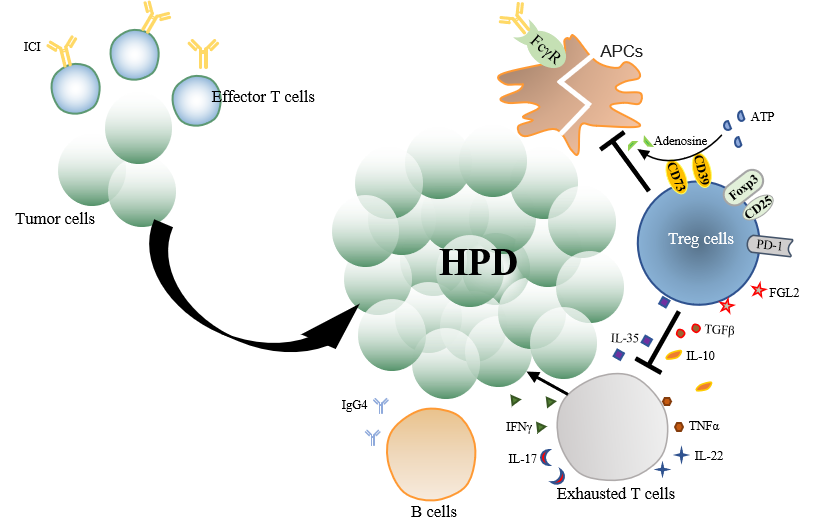
The mechanism of action underlying ICI is the removal of the “braking” function of immune checkpoints and reduction in the escape of tumor cells to enhance the anti-tumor immune response of effector T cells[39]. ICIs reverse the immunosuppressive state of T cells by disrupting the programmed cell death-1/programmed cell death-ligand 1 (PD-1/PD-L1) axis[40]. However, PD-1 receptors are present not only on the surface of T cells but also on the surface of many innate or acquired immune cells, including NK cells, monocytes, macrophages, Treg cells, and B cells[41]. Furthermore, immune cells have varying impacts on PD-1/PD-L1 axis disruption, boosting or inhibiting immune function. In addition, tumor treatment through ICI intervention may also induce changes in the oncogenic pathways of the tumor cells and result in their rapid proliferation and spread[42]. Therefore, HPD may not be triggered by a single factor, but by a series of events that occur simultaneously. Most of the current studies on the molecular mechanisms of HPD focus on the tumor and the tumor microenvironment. In the next sections, we discuss these in detail to facilitate the understanding of the molecular mechanisms underlying ICI-induced HPD. The molecular mechanisms underlying HPD are shown in Table 3.
Mechanisms summary on hyper-progression after immunotherapy
| Tumor cells | Tumor microenvironment | |
| 1. Loss of expression of tumor-associated antigens[43] 2. Impairment of antigen processing and delivery[44] 3. Persistent upregulation of PD-L1 expression on the surface of tumor cells[45] 4. Apoptotic resistance in tumor cells[46,47] 5. Induced dormancy and senescence of tumor cells[48] 6. Tumor cells undergo dedifferentiation and EMT[49] 7. MDM2/MDM4 amplification and EGFR mutation[58] | Treg cells | 1. Competition with conventional T cells for IL-2 via Foxp3[66,136] 2. Secretion of the anti-inflammatory cytokines TGFβ, IL-10, and IL-35[68,69] 3. The dual expression of CD39 and CD73; the CTLA-4-mediated downregulation of CD80 and CD86 on the surface of APCs[71,73] 4. Production of FGL2 to suppress CD8+ T cells and APCs through FcγRIIb[74,137] 5. Express PD-1 receptors 6. A spatial ecological niche dedicated to immunosuppression[76] |
| T cells | 1. Release the cytokines IFNγ[80], IL-17[86,87], IL-22[88,89], TNFα[90,91], and IL-6[92] 2. The combination of multiple cytokines, such as TGFβ and TNFα[80] or IFNγ and TNFα[93] 3. The binding of CD27 receptor to CD70 ligand[94] | |
| B cells | IgG4 competes with IgG1 to bind to Fc receptors on the surface of immune effector cells[107] | |
| Fc receptor | The binding of the Fc region of the anti-PD-1 antibody to the macrophage FcγR[62] | |
Alteration in the tumor cell types following ICI
HPD is a type of primary resistance to immunotherapy, and the mechanism of its occurrence involves alteration in the tumor cell types and the tumor microenvironment. These changes range from enhanced proliferative capacity, invasiveness, and drug resistance of tumor cells to a reduced immunosuppressive capacity in the tumor microenvironment. The tumor cells themselves are altered due to the following reasons: (1) loss of expression of tumor-associated antigens[43]; (2) impairment of antigen processing and delivery, including the loss of human leukocyte antigen expression, failing to deliver tumor antigens to the cell surface[44]; (3) persistent upregulation of PD-L1 expression on the surface of tumor cells, which competes with ICI for binding to
MDM2/MDM4 amplification and EGFR mutation
In 2017, Kato et al.[12] evaluated 155 patients with advanced tumors and found a 3.9% incidence of HPD. Through Next-Generation Sequencing (NGS), murine double minute 2/4 (MDM2/MDM4) amplification was identified in six patients who had TTF < 2 months and two patients were diagnosed with HPD; in 10 other patients, epidermal growth factor receptor (EGFR) mutations were identified. By multivariate analysis, it was found that MDM2/MDM4 amplification and EGFR mutations were associated with TTF < 2 months. The presence of MDM2 amplification and EGFR mutations in patients with HPD were also found in a clinical study by Singavi et al.[50] and Economopoulou et al.[51]. The MDM2 protein encoded by the MDM2 gene is a major negative regulator of the p53 protein. MDM2 can ligate to the p53 protein through the E3 ubiquitin ligase, and the ubiquitinated p53 can be transferred to the cytoplasm and targeted for degradation by the proteasome[52]. Thus, MDM2 amplification can promote tumorigenesis directly or indirectly through the inhibition of p53. In 2018, Kato et al.[53] extended the scope of NGS sequencing to include 102,878 patients with different malignancies and found MDM2 amplification in 3.5% of patients; this was present in a small proportion of patients in most tumor types, and 97.6% of these patients had potentially targetable genomic co-alterations, which suggested that appropriately targeted drugs could be designed to target MDM2 amplification-induced HPD. Fang et al.[54] conducted preclinical studies using the MDM2 inhibitor, APG-115. It acts as an indirect p53 activator, suppresses M2 macrophage polarization, and slows tumor invasion and progression, improving anti-tumor immunity to anti-PD-1 treatment. APG-115-mediated p53 activation promoted anti-tumor immunity in TME regardless of the Trp53 status of the tumor itself. Sahin et al.[55] also used the MDM2 inhibitor AMG-232 in combination with anti-PD-1 antibody therapy to enhance T cell-mediated killing of tumors regardless of PD-L1 expression. Another MDM2 inhibitor, idasanutlin (RG7388), in combination with cytarabine therapy, is the first to enter phase III clinical trials for AML[56,57].
EGFR is the first identified member of the ErbB family and plays an important role in physiological processes, including cell growth, proliferation, and differentiation. EGFR is also involved in tumor development and immunotherapy-related resistance. A meta-analysis involving 21,047 patients from 35 randomized controlled trials indicated that patients with EGFR wild type had significantly prolonged PFS and OS after treatment with ICI, while those with EGFR mutations did not show any improvement[58]. This in part reflected the fact that EGFR mutations are a cause of ICI resistance. The TME in EGFR mutated lung adenocarcinoma was non-inflammatory; interestingly, the non-inflammatory TME had a high infiltration of CD4+ Treg cells. EGFR signaling activates cJun/cJun N-terminal kinase and reduces the level of interferon regulatory factor-1; the former increases CCL22 and thereby recruits CD4+ Treg cells, while the latter reduces the levels of CXCL10 and CCL5 and, in turn, induces CD8+ T cell infiltration[59]. In addition, EGFR can upregulate the number of immunosuppressive receptors and induce the secretion of cytokines with immunosuppressive functions [IL-6, IL-10, and transforming growth factor (TGFβ)] from the TME, which in turn leads to ICI treatment resistance[60]. To some extent, this may explain the occurrence of HPD in patients with EGFR mutations after ICI treatment; however, the exact mechanism of induction needs to be further elucidated. Other somatic mutations and carcinogenic pathways exist in addition to MDM2 amplification and EGFR mutations. Xiong et al.[61] evaluated the mutational and transcriptional characteristics of tumors before and after anti-PD-1 immunotherapy in two patients who acquired HPD. Somatic mutations in recognized cancer genes, including tumor suppressor genes such as TSC2 and VHL, were discovered, as well as transcriptional activation of carcinogenic pathways including IGF-1, ERK/MAPK, PI3K/AKT, and TGFβ.
Treg cells
Treg cells are important for the maintenance of the body’s immune tolerance. The majority of CD4+ Treg cells are produced by the thymus, which accounts for 10% of circulating CD4+ T cells. The major transcription factor is Foxp3, which determines the phenotypic and functional characteristics of Treg cells[62]. In a normal organism, Treg cells negatively regulate immune cells such as effector T cells to prevent autoimmune overload, while, in tumors, Treg cells exhibit different biological functions[63]. Kang et al.[25] found significantly higher FoxP3+ Treg cells in 74 patients with advanced NSCLC who developed HPD and significantly fewer Treg cells in non-HPD patients (P = 0.024). Therefore, PD-1+ Treg cells could be an effective biomarker for the identification of HPD[64]. Previous studies have shown that high Foxp3+ Treg cell infiltration in tumors is significantly associated with poorer OS[65]. Foxp3 is a transcriptional repressor of
Treg cells secrete the anti-inflammatory cytokines TGFβ, IL-10, and IL-35 to deplete conventional T cells[68,69]. TGF, as a Th1 inhibitor, stimulates the TGFβRI/II receptor on conventional T cells to block IFNγ-induced Th1 activation by inhibiting the expression of two essential Th1 transcription factors, T-bet and IFN regulatory factor 1[70]. Indeed, IL-10+ and IL-35+ Treg cells account for a large proportion of tumors. Gene profiles of conventional T cells exposed to these Treg subtypes were analyzed, and it was discovered that T cells depletion was promoted by IL-35+ Treg, but antitumor effects were inhibited by IL-10+ Treg[68].
Treg cells, which have the dual expression of CD39 and CD73, block T cell activation by adenosine triphosphate (ATP) and generate adenosine to inhibit T cells. CD39 and CD73, respectively, hydrolyze ATP/ADP to AMP and AMP to adenosine, leading to a large enrichment of adenosine around Treg cells. Adenosine can induce actin cytoskeleton rearrangement and hence function as a chemoattractant for dendritic cells (DCs), causing DCs to congregate towards Treg cells[71]. Then, with enhanced leukocyte function-associated antigen-1 stability and expression, Treg cells and DCs create a tight aggregate, decreasing the interaction of T cells to DCs[72]. On the other hand, cytotoxic T-lymphocyte-associated protein 4 (CTLA-4) is mediated by Treg cells, resulting in the downregulation of CD80 and CD86 on the surface of antigen-presenting cells (APCs) to restrict the activation of conventional T cells[73]. However, it is uncertain whether the capacity of Treg cells to control CD80 and CD86 is simply dependent on CTLA-4 expression.
Treg cells produce fibrinogen-like protein 2 (FGL2) to suppress CD8+ T cells and APCs through FcγRIIb. FGL2 is considered a signaling molecule for Treg cells as Foxp3 in Treg stimulates the expression of FGL2[74]. The major immunomodulatory effect of FGL2 is mediated via FcγRIIB in APCs. A study has demonstrated that mice lacking the FcγRIIB receptor develop autoimmune glomerulonephritis[75].
Treg cells can also express PD-1 receptors on their surface, and, although blocking the PD-1/PD-L1 axis activates T cells, Treg cells are also highly active, immune function is greatly affected, and anti-tumor immune efficacy is reduced. However, highly activated Treg cells in lymphoid organs resist newly generated anti-tumor T cells, leading to a more attenuated anti-tumor immune effect. These result in uncontrolled tumors, which may lead to HPD. In addition, Murakami et al.[76] reported a spatial ecological niche dedicated to immunosuppression which is formed between CD8+CD39+PD-1+ T cells and Foxp3+PD-1+ Treg cells due to potential interactions between these cells in close proximity following PD-1 blockade in renal cancer. The shift to an immunosuppressive environment is more pronounced in metastatic foci. Anti-CD25 and PD-1 bispecific antibodies are currently used for treatment to deplete Treg cells. Subsequent treatment with anti-PD-1 antibodies may only enhance conventional T cells and CD8+ T cells[77]. Alternatively, adenosine, a product of Treg cells, could be inhibited by combining the anti-PD-1 antibody with adenosine deaminase for its degradation to inosine, thereby reducing cAMP production to weaken the inhibition of conventional T cells and enhance anti-tumor immunity[62]. The possibility of interfering with the systemic immune system is considerably minimized by precisely destroying Treg cells around tumor cells.
T cells
The function of T cell adaptive immunity is to eliminate tumor cells that positively express antigens[78]. However, ICI-enhanced T cell adaptive immune response cannot completely kill tumor cells, as reported in most clinical trials. Even after ICI treatment, adaptive immunity can promote tumor growth directly or indirectly. As a consequence, some researchers believe that enhanced tumor adaptive immune response may be the root cause of HPD in tumor patients after ICI treatment[42]. T cell immune response can induce changes in gene expression of tumor cells, such as a downregulation of tumor surface antigens[79] and upregulation of other immune checkpoint ligands[45]. However, the underlying mechanism of T cell immune response leading to changes in tumor cells remains changes in tumor cells are still not clear.
The cytokine IFNγ released by T cells may explain a part of the problem. IFNγ, a common cytokine, is involved in several cellular changes, including EMT induction[80]. EMT in tumor cells is related to the upregulation of inhibitory checkpoint ligands[81], resistance to cell-mediated cytotoxicity[82], and the production of immunosuppressive effects[83]. Furthermore, IFNγ has the ability to upregulate immune checkpoint ligands[84], inducing tumor cell dormancy, apoptosis[84], and hyperplasia.
The same cytokine can play different roles in different environments, depending on the length of time it acts on tumor cells. For instance, prolonged exposure to IFNγ and low levels of the cytokine have been demonstrated to have pro-tumorigenic effects[85]. Other cytokines, such as IL-17[86,87], IL-22[88,89], tumor necrosis factor α (TNFα)[90,91], and IL-6[92], are involved in tumor promotion. The combination of multiple cytokines may have a greater tumor-promoting effect than a single cytokine; for example, TGFβ1 leads to demethylation of PD-L1 promoter and TNFα leads to the expression of demethylated promoter and co-induces the overexpression of PD-L1[80]. IFNγ and TNFα can co-induce dormancy in tumor cells to promote carcinogenesis[93]. However, there is no clear answer as to which T cell subsets are mainly responsible for the release of these cytokines.
In addition to cytokines, some studies report that the binding of CD27 receptor to CD70 ligand can directly promote proliferation and differentiation of tumor stem cells[94] or T cell exosomes to induce EMT and lead to rapid tumor progression[95]. Many T cell subsets are involved, including CD4+ T cells[96,97], CD8+ T cells[98,99], Th1 cells[100], Th2 cells[101], Th17 cells[102], and Th22 cells[89]. However, the proportion and spatial distribution of tumor cells and effective infiltration of immune cells may be the watershed response of adaptive immunity when the tumor-immunity balance is broken.
Although there are few studies on non-Treg CD4+ T lymphocytes, after ICI treatment, their levels may show an unexpected increase, which can contribute to the occurrence and development of HPD. A prospective study by Arasanz et al.[103] included 70 patients with advanced NSCLC who underwent ICI treatment. Early detection of HPD in NSCLC by monitoring T cell dynamics showed a strong expansion of highly differentiated CD28-CD4+ T lymphocytes (CD4+ THD) between the first and second treatment cycles in HPD patients and a significant stratification among HPD patients, non-HDP patients, and effective patients (median 1.525, 1.000, and 0.9700, respectively, P = 0.0007). As a consequence, the strong expansion of CD28-CD4+ T lymphocytes in peripheral blood during the first treatment cycle could provide an early differential feature of HPD induced by ICI in the treatment of NSCLC. These studies suggest that CD8+ T cells and Treg cells are involved in the occurrence and development of HPD in TME. However, several innate and adaptive immune cells may be swept into this storm.
B cells
PD-1 can also be expressed on the surface of B cells. Some studies have pointed out that anti-PD-1 antibodies can increase the activation, proliferation, and production of inflammatory cytokines in B cells[104]. However, follow-up studies show that the loss of B cells does not seem to have any effect on the efficacy of ICI treatment[105]. The reason for these differences may be due to the existence of different subsets of B cells. The balance among different B cells (resting B cells, activated B cells, Bregs, and other differentiated B cells) determines the ultimate role of B cells in tumor immunity[106]. Humoral immunity may play a role in carcinogenesis. Wang et al.[107] studied the distribution and mechanism of IgG4 secreted by B cells in the tumor model and found that the increase in B lymphocytes containing IgG4 in cancer tissues and the increase in IgG4 concentration in serum were highly correlated with the poor prognoses of patients with esophageal cancer. Using a mouse model, it was verified that IgG4 competes with IgG1 to bind to Fc receptors on the surface of immune effector cells and suppresses classical immune responses such as antibody-dependent cytotoxicity (ADCC), antibody-dependent phagocytosis, and complement-dependent cytotoxicity. Thus, tumor cell growth was indirectly promoted. Interestingly, nivolumab is essentially IgG4 with a stable S228P mutation and significantly promotes the growth of tumors in mice. However, there are only a few studies on the mechanism of B cells participating in HPD after ICI treatment, and these need further validation.
Fc receptor
The binding of the Fc region of the anti-PD-1 antibody to the macrophage FcγR consumes M1 macrophages and stimulates their differentiation to M2-like form. This is another clear mechanism of HPD after ICI treatment in addition to Treg cell-mediated inhibition of anti-tumor immunity leading to HPD[62]. The antibody consists of F(ab’)2 segment bound to the antigen and Fc region bound to FcγR on the surface of immune cells. The binding of the Fc region of IgG antibody to macrophage FcγR triggers the ADCC effect, consumes M1 macrophages and NK cells, and reduces the anti-tumor immune effect[107,108]. Other studies have shown that many M2-PD-L1+ macrophages were observed in the tumor tissues of NSCLC patients with HPD, which could deplete ICI through Fc-FcγR interaction, induce M2-like differentiation of macrophages, and secrete IL-10 to mediate the HPD occurrence[109,110]. The removal of ICI in the Fc region or knockout of FcγR on the surface of macrophages may be a potential research direction for further improvement[111].
CONCLUSION
HPD occurrence is currently a limitation of ICI treatment and represents the storm-like progression of tumors after ICI administration. The mechanism of HPD is similar to a “tug-of-war” between tumor and anti-tumor effects. Intervention through ICI breaks this balance. It leads to the occurrence of HPD if tumor cells are activated and the anti-tumor effect is inhibited. The side effects of chemotherapy cannot be ignored, although the present incidence of HPD in immune combined chemotherapy has been reduced. One day, we hope to usher in the era of “de-chemotherapy”. Then, it would be necessary to face the problem of HPD due to ICI. Hence, the review provides a significant understanding of the current underlying mechanisms for HPD.
DECLARATIONS
AcknowledgmentsWe thank the editors and the reviewers for their useful feedback that improved this paper.
Authors’ contributionsContributed to the conception of the study: Dong X
Wrote the manuscript: Ding P, Wen L
Helped perform the analysis with constructive discussions: Tong F, Zhang R, Huang Y
All authors reviewed and approved the final report.
Availability of data and materialsNot applicable.
Financial support and sponsorshipNone.
Conflicts of interestAll authors declared that there are no conflicts of interest.
Ethical approval and consent to participateNot applicable.
Consent for publicationNot applicable.
Copyright© The Author(s) 2022.
REFERENCES
1. Lahmar J, Mezquita L, Koscielny S, et al. Immune checkpoint inhibitors (IC) induce paradoxical progression in a subset of non-small cell lung cancer (NSCLC). Ann Oncol 2016;27:vi423.
2. Champiat S, Dercle L, Ammari S, et al. Hyperprogressive disease is a new pattern of progression in cancer patients treated by anti-PD-1/PD-L1. Clin Cancer Res 2017;23:1920-8.
3. Socinski M, Creelan B, Horn L, et al. NSCLC, metastatic CheckMate 026: a phase 3 trial of nivolumab vs investigator’s choice (IC) of platinum-based doublet chemotherapy (PT-DC) as first-line therapy for stage iv/recurrent programmed death ligand 1 (PD-L1)-positive NSCLC. Ann Oncol 2016;27:vi577.
4. Peters S, Cappuzzo F, Horn L, et al. OA03.05 analysis of early survival in patients with advanced non-squamous NSCLC treated with nivolumab vs docetaxel in CheckMate 057. J Thorac Oncol 2017;12:S253.
5. Reck M, Schenker M, Lee KH, et al. Nivolumab plus ipilimumab versus chemotherapy as first-line treatment in advanced non-small-cell lung cancer with high tumour mutational burden: patient-reported outcomes results from the randomised, open-label, phase III CheckMate 227 trial. Eur J Cancer 2019;116:137-47.
6. Ferris RL, Blumenschein G Jr, Fayette J, et al. Nivolumab vs investigator’s choice in recurrent or metastatic squamous cell carcinoma of the head and neck: 2-year long-term survival update of CheckMate 141 with analyses by tumor PD-L1 expression. Oral Oncol 2018;81:45-51.
7. Fradet Y, Bellmunt J, Vaughn DJ, et al. Randomized phase III KEYNOTE-045 trial of pembrolizumab versus paclitaxel, docetaxel, or vinflunine in recurrent advanced urothelial cancer: results of >2 years of follow-up. Ann Oncol 2019;30:970-6.
8. Powles T, Durán I, van der Heijden MS, et al. Atezolizumab versus chemotherapy in patients with platinum-treated locally advanced or metastatic urothelial carcinoma (IMvigor211): a multicentre, open-label, phase 3 randomised controlled trial. Lancet 2018;391:748-57.
9. Ferrara R, Mezquita L, Texier M, et al. Hyperprogressive disease in patients with advanced non-small cell lung cancer treated with PD-1/PD-L1 inhibitors or with single-agent chemotherapy. JAMA Oncol 2018;4:1543-52.
10. Mellema WW, Burgers SA, Smit EF. Tumor flare after start of RAF inhibition in KRAS mutated NSCLC: a case report. Lung Cancer 2015;87:201-3.
11. Aoki M, Shoji H, Nagashima K, et al. Hyperprogressive disease during nivolumab or irinotecan treatment in patients with advanced gastric cancer. ESMO Open 2019;4:e000488.
12. Kato S, Goodman A, Walavalkar V, Barkauskas DA, Sharabi A, Kurzrock R. Hyperprogressors after immunotherapy: analysis of genomic alterations associated with accelerated growth rate. Clin Cancer Res 2017;23:4242-50.
13. Saâda-Bouzid E, Defaucheux C, Karabajakian A, et al. Hyperprogression during anti-PD-1/PD-L1 therapy in patients with recurrent and/or metastatic head and neck squamous cell carcinoma. Ann Oncol 2017;28:1605-11.
14. Kim Y, Kim CH, Lee HY, et al. Comprehensive clinical and genetic characterization of hyperprogression based on volumetry in advanced non-small cell lung cancer treated with immune checkpoint inhibitor. J Thorac Oncol 2019;14:1608-18.
15. Kas B, Talbot H, Ferrara R, et al. Clarification of definitions of hyperprogressive disease during immunotherapy for non-small cell lung cancer. JAMA Oncol 2020;6:1039-46.
16. Matos I, Martin-liberal J, Hierro C, et al. Incidence and clinical implications of a new definition of hyperprogression (HPD) with immune checkpoint inhibitors (ICIs) in patients treated in phase 1 (Ph1) trials. JCO 2018;36:3032.
17. Matos I, Martin-Liberal J, García-Ruiz A, et al. Capturing hyperprogressive disease with immune-checkpoint inhibitors using RECIST 1.1 criteria. Clin Cancer Res 2020;26:1846-55.
18. Gomes da Morais AL, de Miguel M, Cardenas JM, Calvo E. Comparison of radiological criteria for hyperprogressive disease in response to immunotherapy. Cancer Treat Rev 2020;91:102116.
19. Abbar B, De Castelbajac V, Gougis P, et al. Definitions, outcomes, and management of hyperprogression in patients with non-small-cell lung cancer treated with immune checkpoint inhibitors. Lung Cancer 2021;152:109-18.
20. Chen S, Gou M, Yan H, et al. Hyperprogressive disease caused by PD-1 inhibitors for the treatment of pan-cancer. Dis Markers 2021;2021:6639366.
21. Kim JY, Lee KH, Kang J, et al. Hyperprogressive disease during anti-PD-1 (PDCD1) / PD-L1 (CD274) therapy: a systematic review and meta-analysis. Cancers (Basel) 2019;11:1699.
22. Park HJ, Kim KW, Won SE, et al. Definition, incidence, and challenges for assessment of hyperprogressive disease during cancer treatment with immune checkpoint inhibitors: a systematic review and meta-analysis. JAMA Netw Open 2021;4:e211136.
23. Kim CG, Kim KH, Pyo KH, et al. Hyperprogressive disease during PD-1/PD-L1 blockade in patients with non-small-cell lung cancer. Ann Oncol 2019;30:1104-13.
24. Kim SH, Choi CM, Lee DH, et al. Clinical outcomes of nivolumab in patients with advanced non-small cell lung cancer in real-world practice, with an emphasis on hyper-progressive disease. J Cancer Res Clin Oncol 2020;146:3025-36.
25. Kang DH, Chung C, Sun P, et al. Circulating regulatory T cells predict efficacy and atypical responses in lung cancer patients treated with PD-1/PD-L1 inhibitors. Cancer Immunol Immunother 2021; doi: 10.1007/s00262-021-03018-y.
26. Chen Y, Hu J, Bu F, Zhang H, Fei K, Zhang P. Clinical characteristics of hyperprogressive disease in NSCLC after treatment with immune checkpoint inhibitor: a systematic review and meta-analysis. BMC Cancer 2020;20:707.
27. Chubachi S, Yasuda H, Irie H, et al. A case of non-small cell lung cancer with possible “disease flare” on nivolumab treatment. Case Rep Oncol Med 2016;2016:1075641.
28. Bernal Vaca L, Mendoza SD, Vergel JC, Rueda X, Bruges R. Hyperprogression in pediatric melanoma metastatic to the breast treated with a checkpoint inhibitor. Cureus 2019;11:e3859.
29. Kanazu M, Edahiro R, Krebe H, et al. Hyperprogressive disease in patients with non-small cell lung cancer treated with nivolumab: a case series. Thorac Cancer 2018;9:1782-7.
30. Liu C, Piao J, Shang Z. Hyperprogressive disease after radiotherapy combined with anti-PD-1 therapy in renal cell carcinoma: a case report and review of the literature. BMC Urol 2021;21:42.
31. Park JH, Chun SH, Lee YG, et al. Hyperprogressive disease and its clinical impact in patients with recurrent and/or metastatic head and neck squamous cell carcinoma treated with immune-checkpoint inhibitors: Korean cancer study group HN 18-12. J Cancer Res Clin Oncol 2020;146:3359-69.
32. Xu Z, Chen L, Zheng L, et al. Hyperprogressive disease in cervical small cell carcinoma treated by immune checkpoint inhibitor. Onco Targets Ther 2019;12:8873-7.
33. Sun D, Liu D, Liu Q, Hou H. Nivolumab induced hyperprogressive disease in advanced esophageal squamous cell carcinoma. Cancer Biol Ther 2020;21:1097-104.
34. Barham W, Guo R, Park SS, Herrmann J, Dong H, Yan Y. Case report: simultaneous hyperprogression and fulminant myocarditis in a patient with advanced melanoma following treatment with immune checkpoint inhibitor therapy. Front Immunol 2020;11:561083.
35. Yang N, Zhang PL, Liu ZJ. Iodine-125 radioactive particles antagonize hyperprogressive disease following immunotherapy: a case report. Medicine (Baltimore) 2020;99:e22933.
36. Peng Y, Zhang L, Zeng T, et al. Characterization of hyperprogression after immunotherapy in a lung adenocarcinoma patient with strong expression of programmed death ligand 1. J Thorac Oncol 2020;15:e4-8.
37. Feng D, Guan Y, Liu M, et al. Excellent response to atezolizumab after clinically defined hyperprogression upon previous treatment with pembrolizumab in metastatic triple-negative breast cancer: a case report and review of the literature. Front Immunol 2021;12:608292.
38. Sama S, Hadfield MJ, Lopetegui Lia N, Vredenburgh J. Hyperprogression in PDL1 expressive, recurrent gastroesophageal-junction adenocarcinoma after pembrolizumab. Cureus 2019;11:e4862.
39. Iwai Y, Hamanishi J, Chamoto K, Honjo T. Cancer immunotherapies targeting the PD-1 signaling pathway. J Biomed Sci 2017;24:26.
40. Pardoll DM. The blockade of immune checkpoints in cancer immunotherapy. Nat Rev Cancer 2012;12:252-64.
41. Keir ME, Butte MJ, Freeman GJ, Sharpe AH. PD-1 and its ligands in tolerance and immunity. Annu Rev Immunol 2008;26:677-704.
42. Marcucci F, Rumio C. The tumor-promoting effects of the adaptive immune system: a cause of hyperprogressive disease in cancer? Cell Mol Life Sci 2021;78:853-65.
43. Kmieciak M, Knutson KL, Dumur CI, Manjili MH. HER-2/neu antigen loss and relapse of mammary carcinoma are actively induced by T cell-mediated anti-tumor immune responses. Eur J Immunol 2007;37:675-85.
44. Facoetti A, Nano R, Zelini P, et al. Human leukocyte antigen and antigen processing machinery component defects in astrocytic tumors. Clin Cancer Res 2005;11:8304-11.
45. Marcucci F, Rumio C, Corti A. Tumor cell-associated immune checkpoint molecules - drivers of malignancy and stemness. Biochim Biophys Acta Rev Cancer 2017;1868:571-83.
46. Breunig C, Pahl J, Küblbeck M, et al. MicroRNA-519a-3p mediates apoptosis resistance in breast cancer cells and their escape from recognition by natural killer cells. Cell Death Dis 2017;8:e2973.
47. Noh KH, Kim SH, Kim JH, et al. API5 confers tumoral immune escape through FGF2-dependent cell survival pathway. Cancer Res 2014;74:3556-66.
48. Brenner E, Schörg BF, Ahmetlić F, et al. Cancer immune control needs senescence induction by interferon-dependent cell cycle regulator pathways in tumours. Nat Commun 2020;11:1335.
49. Romeo E, Caserta CA, Rumio C, Marcucci F. The vicious cross-talk between tumor cells with an EMT phenotype and cells of the immune system. Cells 2019;8:460.
50. Singavi A, Menon S, Kilari D, et al. Predictive biomarkers for hyper-progression (HP) in response to immune checkpoint inhibitors (ICI) - analysis of somatic alterations (SAs). Ann Oncol 2017;28:v405.
51. Economopoulou P, Anastasiou M, Papaxoinis G, et al. Patterns of response to immune checkpoint inhibitors in association with genomic and clinical features in patients with head and neck squamous cell carcinoma (HNSCC). Cancers (Basel) 2021;13:286.
52. Zhao K, Yang Y, Zhang G, et al. Regulation of the Mdm2-p53 pathway by the ubiquitin E3 ligase MARCH7. EMBO Rep 2018;19:305-19.
53. Kato S, Ross JS, Gay L, et al. Analysis of MDM2 amplification: next-generation sequencing of patients with diverse malignancies. JCO Precis Oncol 2018:2018.
54. Fang DD, Tang Q, Kong Y, et al. MDM2 inhibitor APG-115 synergizes with PD-1 blockade through enhancing antitumor immunity in the tumor microenvironment. J Immunother Cancer 2019;7:327.
55. Sahin I, Zhang S, Navaraj A, et al. AMG-232 sensitizes high MDM2-expressing tumor cells to T-cell-mediated killing. Cell Death Discov 2020;6:57.
56. Montesinos P, Beckermann BM, Catalani O, et al. MIRROS: a randomized, placebo-controlled, phase III trial of cytarabine ± idasanutlin in relapsed or refractory acute myeloid leukemia. Future Oncol 2020;16:807-15.
57. Konopleva M, Martinelli G, Daver N, et al. MDM2 inhibition: an important step forward in cancer therapy. Leukemia 2020;34:2858-74.
58. Sun L, Zhang L, Yu J, et al. Clinical efficacy and safety of anti-PD-1/PD-L1 inhibitors for the treatment of advanced or metastatic cancer: a systematic review and meta-analysis. Sci Rep 2020;10:2083.
59. Sugiyama E, Togashi Y, Takeuchi Y, et al. Blockade of EGFR improves responsiveness to PD-1 blockade in EGFR-mutated non-small cell lung cancer. Sci Immunol 2020;5:eaav3937.
60. Akbay EA, Koyama S, Carretero J, et al. Activation of the PD-1 pathway contributes to immune escape in EGFR-driven lung tumors. Cancer Discov 2013;3:1355-63.
61. Xiong D, Wang Y, Singavi AK, Mackinnon AC, George B, You M. Immunogenomic landscape contributes to hyperprogressive disease after anti-PD-1 immunotherapy for cancer. iScience 2018;9:258-77.
62. Tay C, Qian Y, Sakaguchi S. Hyper-progressive disease: the potential role and consequences of T-regulatory cells foiling anti-PD-1 cancer immunotherapy. Cancers (Basel) 2020;13:48.
63. Gallimore A, Quezada SA, Roychoudhuri R. Regulatory T cells in cancer: where are we now? Immunology 2019;157:187-9.
64. Kamada T, Togashi Y, Tay C, et al. PD-1+ regulatory T cells amplified by PD-1 blockade promote hyperprogression of cancer. Proc Natl Acad Sci U S A 2019;116:9999-10008.
65. Shang B, Liu Y, Jiang SJ, Liu Y. Prognostic value of tumor-infiltrating FoxP3+ regulatory T cells in cancers: a systematic review and meta-analysis. Sci Rep 2015;5:15179.
66. Ono M, Yaguchi H, Ohkura N, et al. Foxp3 controls regulatory T-cell function by interacting with AML1/Runx1. Nature 2007;446:685-9.
67. Wu Y, Borde M, Heissmeyer V, et al. FOXP3 controls regulatory T cell function through cooperation with NFAT. Cell 2006;126:375-87.
68. Sawant DV, Yano H, Chikina M, et al. Adaptive plasticity of IL-10+ and IL-35+ Treg cells cooperatively promotes tumor T cell exhaustion. Nat Immunol 2019;20:724-35.
69. Turnis ME, Sawant DV, Szymczak-Workman AL, et al. Interleukin-35 limits anti-tumor immunity. Immunity 2016;44:316-29.
70. Lin JT, Martin SL, Xia L, Gorham JD. TGF-beta 1 uses distinct mechanisms to inhibit IFN-gamma expression in CD4+ T cells at priming and at recall: differential involvement of Stat4 and T-bet. J Immunol 2005;174:5950-8.
71. Ring S, Pushkarevskaya A, Schild H, et al. Regulatory T cell-derived adenosine induces dendritic cell migration through the Epac-Rap1 pathway. J Immunol 2015;194:3735-44.
72. Onishi Y, Fehervari Z, Yamaguchi T, Sakaguchi S. Foxp3+ natural regulatory T cells preferentially form aggregates on dendritic cells in vitro and actively inhibit their maturation. Proc Natl Acad Sci U S A 2008;105:10113-8.
73. Qureshi OS, Zheng Y, Nakamura K, et al. Trans-endocytosis of CD80 and CD86: a molecular basis for the cell-extrinsic function of CTLA-4. Science 2011;332:600-3.
74. Gavin MA, Rasmussen JP, Fontenot JD, et al. Foxp3-dependent programme of regulatory T-cell differentiation. Nature 2007;445:771-5.
75. Boross P, Arandhara VL, Martin-Ramirez J, et al. The inhibiting Fc receptor for IgG, FcγRIIB, is a modifier of autoimmune susceptibility. J Immunol 2011;187:1304-13.
76. Murakami T, Tanaka N, Takamatsu K, et al. Multiplexed single-cell pathology reveals the association of CD8 T-cell heterogeneity with prognostic outcomes in renal cell carcinoma. Cancer Immunol Immunother 2021;70:3001-13.
77. Arce Vargas F, Furness AJS, Solomon I, et al; Melanoma TRACERx Consortium, Renal TRACERx Consortium, Lung TRACERx Consortium. Fc-optimized anti-CD25 depletes tumor-infiltrating regulatory T cells and synergizes with PD-1 blockade to eradicate established tumors. Immunity 2017;46:577-86.
78. Boisgerault N, Kottke T, Pulido J, et al. Functional cloning of recurrence-specific antigens identifies molecular targets to treat tumor relapse. Mol Ther 2013;21:1507-16.
79. Matsushita H, Vesely MD, Koboldt DC, et al. Cancer exome analysis reveals a T-cell-dependent mechanism of cancer immunoediting. Nature 2012;482:400-4.
80. Asgarova A, Asgarov K, Godet Y, et al. PD-L1 expression is regulated by both DNA methylation and NF-kB during EMT signaling in non-small cell lung carcinoma. Oncoimmunology 2018;7:e1423170.
81. Prestipino A, Zeiser R. Clinical implications of tumor-intrinsic mechanisms regulating PD-L1. Sci Transl Med 2019;11:eaav4810.
82. Terry S, Buart S, Tan TZ, et al. Acquisition of tumor cell phenotypic diversity along the EMT spectrum under hypoxic pressure: consequences on susceptibility to cell-mediated cytotoxicity. Oncoimmunology 2017;6:e1271858.
83. Terry S, Savagner P, Ortiz-Cuaran S, et al. New insights into the role of EMT in tumor immune escape. Mol Oncol 2017;11:824-46.
84. Benci JL, Xu B, Qiu Y, et al. Tumor interferon signaling regulates a multigenic resistance program to immune checkpoint blockade. Cell 2016;167:1540-54.e12.
85. Minn AJ, Wherry EJ. Combination cancer therapies with immune checkpoint blockade: convergence on interferon signaling. Cell 2016;165:272-5.
86. Zhang Y, Zoltan M, Riquelme E, et al. Immune cell production of interleukin 17 induces stem cell features of pancreatic intraepithelial neoplasia cells. Gastroenterology 2018;155:210-23.e3.
87. Guo N, Shen G, Zhang Y, Moustafa AA, Ge D, You Z. Interleukin-17 promotes migration and invasion of human cancer cells through upregulation of MTA1 expression. Front Oncol 2019;9:546.
88. Kryczek I, Lin Y, Nagarsheth N, et al. IL-22(+)CD4(+) T cells promote colorectal cancer stemness via STAT3 transcription factor activation and induction of the methyltransferase DOT1L. Immunity 2014;40:772-84.
89. Khosravi N, Caetano MS, Cumpian AM, et al. IL22 promotes Kras-mutant lung cancer by induction of a protumor immune response and protection of stemness properties. Cancer Immunol Res 2018;6:788-97.
90. Landsberg J, Kohlmeyer J, Renn M, et al. Melanomas resist T-cell therapy through inflammation-induced reversible dedifferentiation. Nature 2012;490:412-6.
91. Bertrand F, Montfort A, Marcheteau E, et al. TNFα blockade overcomes resistance to anti-PD-1 in experimental melanoma. Nat Commun 2017;8:2256.
92. Chen L, Heymach JV, Qin FX, Gibbons DL. The mutually regulatory loop of epithelial-mesenchymal transition and immunosuppression in cancer progression. Oncoimmunology 2015;4:e1002731.
93. Müller-Hermelink N, Braumüller H, Pichler B, et al. TNFR1 signaling and IFN-gamma signaling determine whether T cells induce tumor dormancy or promote multistage carcinogenesis. Cancer Cell 2008;13:507-18.
94. Schürch C, Riether C, Matter MS, Tzankov A, Ochsenbein AF. CD27 signaling on chronic myelogenous leukemia stem cells activates Wnt target genes and promotes disease progression. J Clin Invest 2012;122:624-38.
95. Min H, Sun X, Yang X, et al. Exosomes derived from irradiated esophageal carcinoma-infiltrating T cells promote metastasis by inducing the epithelial-mesenchymal transition in esophageal cancer cells. Pathol Oncol Res 2018;24:11-8.
96. Goebel L, Grage-Griebenow E, Gorys A, et al. CD4+ T cells potently induce epithelial-mesenchymal-transition in premalignant and malignant pancreatic ductal epithelial cells-novel implications of CD4+ T cells in pancreatic cancer development. Oncoimmunology 2015;4:e1000083.
97. Aspord C, Pedroza-Gonzalez A, Gallegos M, et al. Breast cancer instructs dendritic cells to prime interleukin 13-secreting CD4+ T cells that facilitate tumor development. J Exp Med 2007;204:1037-47.
98. Schürch C, Riether C, Amrein MA, Ochsenbein AF. Cytotoxic T cells induce proliferation of chronic myeloid leukemia stem cells by secreting interferon-γ. J Exp Med 2013;210:605-21.
99. Sanchez-Perez L, Kottke T, Diaz RM, et al. Potent selection of antigen loss variants of B16 melanoma following inflammatory killing of melanocytes in vivo. Cancer Res 2005;65:2009-17.
100. Sosa MS, Bragado P, Aguirre-Ghiso JA. Mechanisms of disseminated cancer cell dormancy: an awakening field. Nat Rev Cancer 2014;14:611-22.
101. Ochi A, Nguyen AH, Bedrosian AS, et al. MyD88 inhibition amplifies dendritic cell capacity to promote pancreatic carcinogenesis via Th2 cells. J Exp Med 2012;209:1671-87.
102. McAllister F, Bailey JM, Alsina J, et al. Oncogenic Kras activates a hematopoietic-to-epithelial IL-17 signaling axis in preinvasive pancreatic neoplasia. Cancer Cell 2014;25:621-37.
103. Arasanz H, Zuazo M, Bocanegra A, et al. Early detection of hyperprogressive disease in non-small cell lung cancer by monitoring of systemic T cell dynamics. Cancers (Basel) 2020;12:344.
104. Thibult ML, Mamessier E, Gertner-Dardenne J, et al. PD-1 is a novel regulator of human B-cell activation. Int Immunol 2013;25:129-37.
105. Damsky W, Jilaveanu L, Turner N, et al. B cell depletion or absence does not impede anti-tumor activity of PD-1 inhibitors. J Immunother Cancer 2019;7:153.
106. Zhao KL, Yang XJ, Jin HZ, Zhao L, Hu JL, Qin WJ. Double-edge role of B cells in tumor immunity: potential molecular mechanism. Curr Med Sci 2019;39:685-9.
107. Wang H, Xu Q, Zhao C, et al. An immune evasion mechanism with IgG4 playing an essential role in cancer and implication for immunotherapy. J Immunother Cancer 2020;8:e000661.
108. Karagiannis P, Gilbert AE, Josephs DH, et al. IgG4 subclass antibodies impair antitumor immunity in melanoma. J Clin Invest 2013;123:1457-74.
109. Andreu P, Johansson M, Affara NI, et al. FcRgamma activation regulates inflammation-associated squamous carcinogenesis. Cancer Cell 2010;17:121-34.
110. Kim SR, Chun SH, Kim JR, et al. The implications of clinical risk factors, CAR index, and compositional changes of immune cells on hyperprogressive disease in non-small cell lung cancer patients receiving immunotherapy. BMC Cancer 2021;21:19.
111. Lo Russo G, Moro M, Sommariva M, et al. Antibody-Fc/FcR interaction on macrophages as a mechanism for hyperprogressive disease in non-small cell lung cancer subsequent to PD-1/PD-L1 blockade. Clin Cancer Res 2019;25:989-99.
112. Costantini A, Fallet V, Corny J, et al. Nivolumab-refractory patients with advanced non-small-cell lung cancer. Lung Cancer 2019;130:128-34.
113. Matsuo N, Azuma K, Kojima T, et al. Comparative incidence of immune-related adverse events and hyperprogressive disease in patients with non-small cell lung cancer receiving immune checkpoint inhibitors with and without chemotherapy. Invest New Drugs 2021;39:1150-8.
114. Schuiveling M, Tonk EHJ, Verheijden RJ, Suijkerbuijk KPM. Hyperprogressive disease rarely occurs during checkpoint inhibitor treatment for advanced melanoma. Cancer Immunol Immunother 2021;70:1491-6.
115. Hagi T, Kurokawa Y, Kawabata R, et al. Multicentre biomarker cohort study on the efficacy of nivolumab treatment for gastric cancer. Br J Cancer 2020;123:965-72.
116. Kim CG, Kim C, Yoon SE, et al. Hyperprogressive disease during PD-1 blockade in patients with advanced hepatocellular carcinoma. J Hepatol 2021;74:350-9.
117. Hwang I, Park I, Yoon SK, Lee JL. Hyperprogressive disease in patients with urothelial carcinoma or renal cell carcinoma treated with PD-1/PD-L1 inhibitors. Clin Genitourin Cancer 2020;18:e122-33.
118. Rodriguez Freixinos V, Garcia A, Fasani R, et al. Immune profile and outcomes of patients (pts) with gynecological malignancies (GYN) enrolled in early phases immunotherapy (IO) trials. JCO 2018;36:5595.
119. Chiba Y, Kawanami T, Yamasaki K, Uchimura K, Matsuyama A, Yatera K. Hyper-progressive disease after immune checkpoint inhibitor in SMARCA4-deficient small-cell lung carcinoma. Respirol Case Rep 2020;8:e00667.
120. Fricke J, Mambetsariev I, Pharaon R, Subbiah S, Rajurkar S, Salgia R. Hyperprogression on immunotherapy with complete response to chemotherapy in a NSCLC patient with high PD-L1 and STK11: a case report. Medicine (Baltimore) 2020;99:e22323.
121. Martorana F, Lanzafame K, Pavone G, et al. Early gastrointestinal progression to immunotherapy in lung cancer: a report of two cases. Case Rep Oncol Med 2021;2021:6692538.
122. Lin Z, Liu Q, Wei Q, Lin L, Chen X, Xue D. Hyperprogressive disease in advanced cancer patients with liver metastasis treated with PD-1 inhibitors: two case reports. Ann Transl Med 2020;8:1100.
123. Oguri T, Sasada S, Seki S, et al. A case of hyperprogressive disease following atezolizumab therapy for pulmonary pleomorphic carcinoma with epidermal growth factor receptor mutation. Respir Med Case Rep 2021;33:101405.
124. Wang W, Wu M, Liu M, et al. Hyperprogression to camrelizumab in a patient with esophageal squamous cell carcinoma harboring EGFR kinase domain duplication. J Immunother Cancer 2020;8:e000793.
125. Huang LT, Ma JT, Zhang SL, et al. Durable clinical response to pyrotinib after resistance to prior anti-HER2 therapy for HER2-positive advanced gastric cancer: a case report. Front Oncol 2019;9:1453.
126. Singh B, Kaur P, Maroules M. Hyperprogression in a patient with hepatocellular cancer treated with atezolizumab and bevacizumab: a case report and review of literature. J Investig Med High Impact Case Rep 2021;9:2324709621992207.
127. Wong DJ, Lee J, Choo SP, Thng CH, Hennedige T. Hyperprogressive disease in hepatocellular carcinoma with immune checkpoint inhibitor use: a case series. Immunotherapy 2019;11:167-75.
128. Chan KH, Lakkasani S, Ramahi A, Shaaban HS. Hyperprogressive disease in an advanced stage colon cancer patient on pembrolizumab: a case report. Cureus 2020;12:e7764.
129. Yilmaz M, Akovali B. Hyperprogression after nivolumab for melanoma: a case report. J Oncol Pharm Pract 2020;26:244-51.
130. Forschner A, Niessner H, Möller Y, et al. Genomics of immunotherapy-associated hyperprogressors-letter. Clin Cancer Res 2017;23:6374-5.
131. Koukourakis IM, Kouloulias V, Koukourakis MI. Radio-immunotherapy: a case report of ‘abscopal hyper-progression’? Cureus 2020;12:e10117.
132. Grecea M, Marabelle A, Ammari S, Massard C, Champiat S. Managing hyperprogressive disease in the era of programmed cell death protein 1/programmed death-ligand 1 blockade: a case discussion and review of the literature. Oncologist 2020;25:369-74.
133. Ikushima H, Sakatani T, Ohara S, et al. Cisplatin plus pemetrexed therapy and subsequent immune checkpoint inhibitor administration for malignant peritoneal mesothelioma without pleural lesions: case report. Medicine (Baltimore) 2020;99:e19956.
134. Xiang JJ, Uy NF, Minja FJ, Verter EE, Burtness BA. Hyperprogression after one dose of nivolumab in sinonasal cancer: a case report. Laryngoscope 2020;130:907-10.
135. Chan AS, Ng VY, Snider J, Kallen ME, Miller KD. Hyperprogression of liver metastasis with neoadjuvant immunotherapy for soft tissue sarcoma. Cureus 2020;12:e8575.
136. Pandiyan P, Zheng L, Ishihara S, Reed J, Lenardo MJ. CD4+CD25+Foxp3+ regulatory T cells induce cytokine deprivation-mediated apoptosis of effector CD4+ T cells. Nat Immunol 2007;8:1353-62.
Cite This Article
Export citation file: BibTeX | RIS
OAE Style
Ding P, Wen L, Tong F, Zhang R, Huang Y, Dong X. Mechanism underlying the immune checkpoint inhibitor-induced hyper-progressive state of cancer. Cancer Drug Resist 2022;5:147-64. http://dx.doi.org/10.20517/cdr.2021.104
AMA Style
Ding P, Wen L, Tong F, Zhang R, Huang Y, Dong X. Mechanism underlying the immune checkpoint inhibitor-induced hyper-progressive state of cancer. Cancer Drug Resistance. 2022; 5(1): 147-64. http://dx.doi.org/10.20517/cdr.2021.104
Chicago/Turabian Style
Ding, Peng, Lu Wen, Fan Tong, Ruiguang Zhang, Yu Huang, Xiaorong Dong. 2022. "Mechanism underlying the immune checkpoint inhibitor-induced hyper-progressive state of cancer" Cancer Drug Resistance. 5, no.1: 147-64. http://dx.doi.org/10.20517/cdr.2021.104
ACS Style
Ding, P.; Wen L.; Tong F.; Zhang R.; Huang Y.; Dong X. Mechanism underlying the immune checkpoint inhibitor-induced hyper-progressive state of cancer. Cancer Drug Resist. 2022, 5, 147-64. http://dx.doi.org/10.20517/cdr.2021.104
About This Article
Special Issue
Copyright

Data & Comments
Data


 Cite This Article 9 clicks

 Like This Article 5
likes
Like This Article 5
likes


Comments
Comments must be written in English. Spam, offensive content, impersonation, and private information will not be permitted. If any comment is reported and identified as inappropriate content by OAE staff, the comment will be removed without notice. If you have any queries or need any help, please contact us at support@oaepublish.com.